


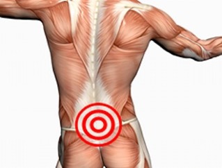
Lumbar osteochondrosisis one of the most common diseases of the spine.
It is characterized by deformation of the cartilage tissue of the vertebrae.
The spine remains flexible and agile as long as the vertebrae are healthy.
If the condition worsens, the intervertebral discs lose their elasticity and begin to dry out.
As a result, patients experience severe pain in the lumbar region.
What's this?
When dystrophic-degenerative changes appear in the tissues of the vertebrae, they gradually begin to collapse. If the vertebrae in the lumbar region suffer primarily, osteochondrosis of the lumbar spine is diagnosed.
Clinical presentation
In osteochondrosis, the cartilage tissue of the vertebrae begins to lose moisture, the elasticity of the intervertebral discs deteriorates. Over time, the height between the vertebrae decreases. Under increased load, the fibrous ring begins to crack, with the intervertebral disc protruding.This leads to tightening of nerve endings and the appearance of pain.
Osteochondrosis progresses without proper treatment. Intervertebral discs harden, their damping properties deteriorate. Increases develop in the bone tissue of the vertebrae, which suppress nerve endings. Because of this, patients cause constant pain.
Degrees and Classifications
Experts distinguish 4 degrees of spinal osteochondrosis:
- The process of destruction of the intervertebral discs begins, the symptoms of the disease are mild, burning sensation, tingling, pain occurs only after physical exertion. Patients talk about the appearance of dull back pain, sometimes radiating to the buttocks.
- The distance between the vertebrae gradually decreases and the annulus fibrosus collapses. Intervertebral discs compress, go beyond physiological boundaries, there is pressure on the nerve roots. Patients complain of tangible pain in the buttocks, thighs, and legs during walking. In addition to pain, a burning sensation, coldness is possible.
- The fibrous rings are destroyed, and the examination reveals intervertebral hernia in the patients. The pain is constantly disturbing regardless of the load.
- Bone outgrowths of the vertebrae are visible. Cartilage withers, the patient may have difficulty moving. As a result, the lumbar spine loses its mobility and flexibility and the patient becomes disabled.
| Osteochondrosis is also classified according to the type of disease course: | |
|
|
Experts distinguish this period of osteochondrosis:
- debut;
- exacerbation;
- remission;
- stabilization.
Treatment is chosen depending on the stage of the disease, the nature of the deformity and the severity of the symptoms of the disease.
ICD 10 code
According to the International Classification of Diseases, osteochondrosis of the spine was coded M42. There are juvenile (M42. 0), adult (M42. 1) and unspecified (M42. 9) osteochondrosis separately.
Prevalence and Significance
The lumbar region is more prone to osteochondrosis than the rest of the spine. This is due to the increased load in this area because you have to bear the weight. In the case of a weak muscle ligament, the condition of the intervertebral discs begins to deteriorate rapidly and is destroyed.
Most often, people who have crossed the 30-year limitsuffer from lumbar spine osteochondrosis. Although found in younger patients. Pain in the lumbar region can be diagnosed with osteochondrosis in nearly 80% of physicians with complaints.
Studies in patients over 40 years of age have shown that most of them have characteristic changes in the intervertebral discs. But in the absence of clinical manifestations, a person cannot be considered sick.
In the absence of proper therapy, the disease progresses. It leads to disability in neglected forms.
Risk Factors and Causes
Representatives of such professions often encounter osteochondrosis: programmers, office workers, builders, movers, waiters, drivers.
Risk factors that increase the likelihood of developing osteochondrosis:
- overweight;
- unhealthy diet;
- posture problems;
- genetic predisposition;
- lack of sleep;
- common stress;
- persistent hypothermia;
- should be in an awkward position for a long time;
- low physical activity.
Causes of lumbar spine osteochondrosis include::
- the natural aging processes of the body;
- metabolic problems;
- back injuries;
- back problems;
- flat feet;
- intense physical activity such as weight lifting;
- problems with the joints of the spine (rheumatoid arthritis);
- endocrine diseases;
- digestive and cardiovascular problems.
Some experts believe that the predisposition to osteochondrosis spreads at the genetic level.
Consequences
A change in cartilage tissue between the vertebraeleads to deterioration of the annulus fibrosus and the appearance of a hernia. Patients begin to complain of severe pain in the lumbar region that radiates to the buttocks, thighs, and lower legs. But this is not the only possible complication in osteochondrosis.
Prolonged irritation of the spinal nerve leads to inflammation.Sciatica is lumbar in patients.
Osteochondrosis can lead tosciatica(inflammation of the sciatic nerve). The disease leads to severe pain in the lower back, numbness of the legs. Patients begin to walk, leaning to one side. This causes further curvature of the spine and further destruction of the intervertebral discs.
Osteochondrosis provokes vertebral instability. The lumbar region begins to move from the sacrum under the influence of body weight. In women, such instability triggers the appearance of problems with the internal organs (the uterus, ovaries, appendages suffer), in men - by force.
When the intervertebral discs are destroyed, the blood supply to the spinal cord is interrupted, the movement of the vertebrae leads to compression myelopathy.
Cauda equina syndrome is considered to be the most dangerous complication. It is due to the fact that the nerve roots are affected. In severe cases, osteochondrosis causes paresis of the lower extremities or paralysis of both feet.
Adverse effects can be prevented by seeking medical attention at the first appearance of symptoms and not ignoring the need for treatment.
Symptoms
Osteochondrosis does not appear immediately. In the initial stage, the patient has no pain or discomfort. Complaints usually occur when the disease progresses to stage 2.
Main symptoms of lumbar osteochondrosis::
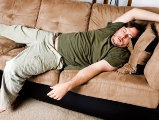
- lumbar pain that worsens as the disease progresses;
- disabled: problems occur when trying to bend down, turn around, feelings when changing body position are described by patients as an “electric shock” while many feel pain in their legs;
- changes in the sensitivity of the limbs, which occurred in the background of damage to the nerve roots, burning sensation, numbness, crawling, tingling in the affected area;
- muscle weakness, lack of tendon reflexes;
- local temperature drop;
- increased sweating;
- pale, dry skin in the problem area;
- urinary disorders, sexual dysfunction (in severe osteochondrosis).
Some patients have cramps in the arteries of the legs. But symptoms are expressed only in the acute form of osteochondrosis. Exacerbation may begin abruptly with hypothermia, awkward movement, or after intense physical activity.
Which doctor is treating you?
If you have back pain,consult an orthopedist and neurologist. The test assesses the patient's neurological condition, checking how the spine performs its functions. Doctors also assess the condition of the back and buttocks.
For experienced professionals, one test is sufficient to make a preliminary diagnosis. But to confirm this, the patient is sent to hardware diagnostics.
Diagnostic Methods
The simplest and most readily available method for detecting osteochondrosis isradiography. Computer or magnetic resonance imaging is required for a more accurate image.
MRIallows you to examine the condition of your spine as accurately as possible. Indeed, during the process, images of the problem area are taken layer by layer.
Management

Therapy tactics are selected by the physician depending on the patient's condition, the stage of osteochondrosis, and the clinical manifestation of the disease.
Your doctor may prescribe:
- drug therapy, non-steroidal anti-inflammatory drugs, hormonal drugs, analgesics are chosen;
- drug blockade, painkillers, hormonal drugs are injected into the affected area or into the muscles around the problematic vertebra, which almost immediately relieves inflammation and removes pain;
- hand therapy, massage, physiotherapyis recommended after stopping the acute stage of the disease, physiotherapy can increase the effectiveness of medication;
- medical gymnastics;
- acupuncture.
Action required in advanced cases. Surgery is prescribed in situations where conservative treatment does not produce the expected results.
Conclusion
As thedystrophic-degenerative changes progress, osteochondrosis is diagnosed in the cartilage tissues of the lumbar spine. In advanced forms, this disease can not only lead to the appearance of permanent severe pain, but can also cause paresis, paralysis of the lower extremities.
- You may suspect osteochondrosis with lower back pain. As the disease progresses, the pain increases significantly, and the lower back loses its mobility.
- Depending on the degree ofdestruction of the intervertebral discs, the disease has 4 stages.
- This diagnosis is more common after the age of 30. Almost 80% of patients who see a doctor for back pain are diagnosed with osteochondrosis.
- People who lead an inactive lifestyle are prone to osteochondrosis, are in an unnatural state for a long time, and often experience physical overload.
- The main symptoms of osteochondrosis are pain and low back movement.
- Patients have foot problems due to the destruction of the intervertebral discs of the lumbar spine.
- If left untreated, pain may increase, sciatica, spinal instability, and compression myelopathy may develop. In advanced cases, it paralyzes the lower limbs.
- In case of pain,consult a neurologist and orthopedist. The patient is referred for radiography, computed tomography, or magnetic resonance imaging.
- Depending on the condition,prescribes medication, blockages, massage, hand therapy, physiotherapy, physiotherapy exercises, or surgeries.